
Dopamine is not just a neurotransmitter but also a molecule involved in immune cell interaction, which improves T-B-cell immune synapse in human lymph nodes.
Viewpoint on:
Papa, I., Saliba, D., Ponzoni, M. et al., TFH-derived dopamine accelerates productive synapses in germinal centres. Nature 547, 318–323 (2017). https://doi.org/10.1038/nature23013
The starting point
Dopamine is one of the best-studied neurotransmitters in the brain. Despite a body of evidence suggesting a functional role of dopamine on inflammation, studies describing the role of dopamine in immune cells appeared only in the 1990es. Nevertheless, since then many studies have been published (for recent reviews, see[1-3]).
Nowadays, it is well described that immune cells are not just able to respond to external dopamine, but that many, if not all of them, can synthesize it as well. Bergquist et al. described in the 1990s the presence of catecholamines in immune cells, thus suggesting an autocrine loop on immune function[4]. These findings were later confirmed by Musso et al. [5].
Only few years later, Marino et al. [6] demonstrated that PBMCs can synthesize, release and degrade catecholamines. Later, catecholamine synthesis by immune cells was also demonstrated in different lymphoid organs, with lymph nodes showing the highest and thymus the lowest concentration [7].
Despite these interesting findings, it is still not yet fully understood how immune cells use the dopaminergic pathway for autocrine and paracrine communication.
Considering the fact that the nervous system and the immune system share signaling pathways which were previously considered to be system-specific, and due to the similarity of the immune and neuronal synapses, Papa et al. investigated the possible presence of neuronal signaling molecules in immune cells in lymph nodes, and their possible role in the immune cell communication.
The discovery
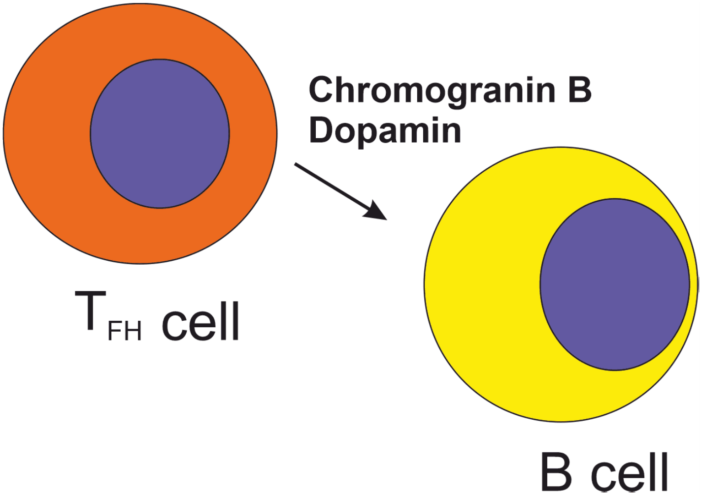
The results nicely show the presence of Chromogranin-B (CgB), a protein expressed in dense-core neuroendocrine secretory granules containing catecholamines, in T follicular helper (TFH) cells in human lymph nodes. CgB expression increases during TFH maturation, indicating a rapid, post-transcriptional regulation. CgB was altered in pathologic conditions, thus suggesting a functional role of neuroendocrine secretory granules in TFH cells. Tyrosine hydroxylase (TH), the rate-limiting enzyme required for catecholamine synthesis, was also present in TFH, as well as dopamine-beta-hydroxylase (DBH), which converts dopamine into norepinephrine. However, only dopamine was detected at high concentrations and was restricted to CgB+ TFH cells.
The released dopamine was then captured by allogenic germinal centre B cells. Inhibition of LFA1/ICAM1 interactions prevented dopamine release, implying that this pathway is required for the dopamine signaling. Papa et al. also showed that dopamine led to a rapid translocation of ICOSL to the surface of germinal centre B cells, and ICOSL triggered then the increase of CD40L on the surface of TFH cells. The authors speculate that dopamine improved the time required by B cells to engage with T cells, thus improving the output without changing B cell fate.
Another important finding of the study was the absence of these dopamine-mediated signaling in the mice, thus suggesting that the dopaminergic signaling pathway is not conserved. Indeed, DBH expression was limited to B cells and dopamine expression in TFH cells was very limited. Also, dopamine did not induce ICOSL translocation to the cell surface in mice.
These results demonstrate that the translation of knowledge from the mouse model to human cells is not always possible.
Discussion
This work nicely described how immune cells use self-synthesized dopamine to interact and modulate immune cell function. The alteration of dopaminergic pathway in pathologic conditions also suggests the dynamic use of the dopaminergic pathway by immune cells.
These findings have many possible clinical consequences. First of all, they propose that the modulation of dopaminergic pathway can be used as a therapeutic target to regulate immune cell functions. On the other hand, clinicians should be aware that dopaminergic drugs might exert side effects on immune cell functions, such as vaccine efficiency. Such possible side effects were already described, but the direct link between neurological therapies and immune response was not taken into consideration.
The absence of comparable dopaminergic signaling in mice also highlights that not all pathways are conserved in mice and humans, and dopaminergic signaling requires research in human models or in humanized animal models for clinical relevance.
References
- Feng Y, Lu Y: Immunomodulatory Effects of Dopamine in Inflammatory Diseases. Front Immunol 2021, 12:663102.
- Ugalde V, Contreras F, Prado C, Chovar O, Espinoza A, Pacheco R: Dopaminergic signalling limits suppressive activity and gut homing of regulatory T cells upon intestinal inflammation. Mucosal Immunol 2021, 14(3):652-666.
- Wang H, Kang R, Tang D: Dopamine as an endogenous regulator of innate immunity in sepsis. Front Immunol 2025, 16:1625368.
- Bergquist J, Tarkowski A, Ekman R, Ewing A: Discovery of endogenous catecholamines in lymphocytes and evidence for catecholamine regulation of lymphocyte function via an autocrine loop. Proc Natl Acad Sci U S A 1994, 91(26):12912-12916.
- Musso NR, Brenci S, Setti M, Indiveri F, Lotti G: Catecholamine content and in vitro catecholamine synthesis in peripheral human lymphocytes. J Clin Endocrinol Metab 1996, 81(10):3553-3557.
- Marino F, Cosentino M, Bombelli R, Ferrari M, Lecchini S, Frigo G: Endogenous catecholamine synthesis, metabolism storage, and uptake in human peripheral blood mononuclear cells. Exp Hematol 1999, 27(3):489-495.
- Qiu YH, Peng YP, Jiang JM, Wang JJ: Expression of tyrosine hydroxylase in lymphocytes and effect of endogenous catecholamines on lymphocyte function. Neuroimmunomodulation 2004, 11(2):75-83.
(Featured image declaration: created by Prof. Rainer Straub)