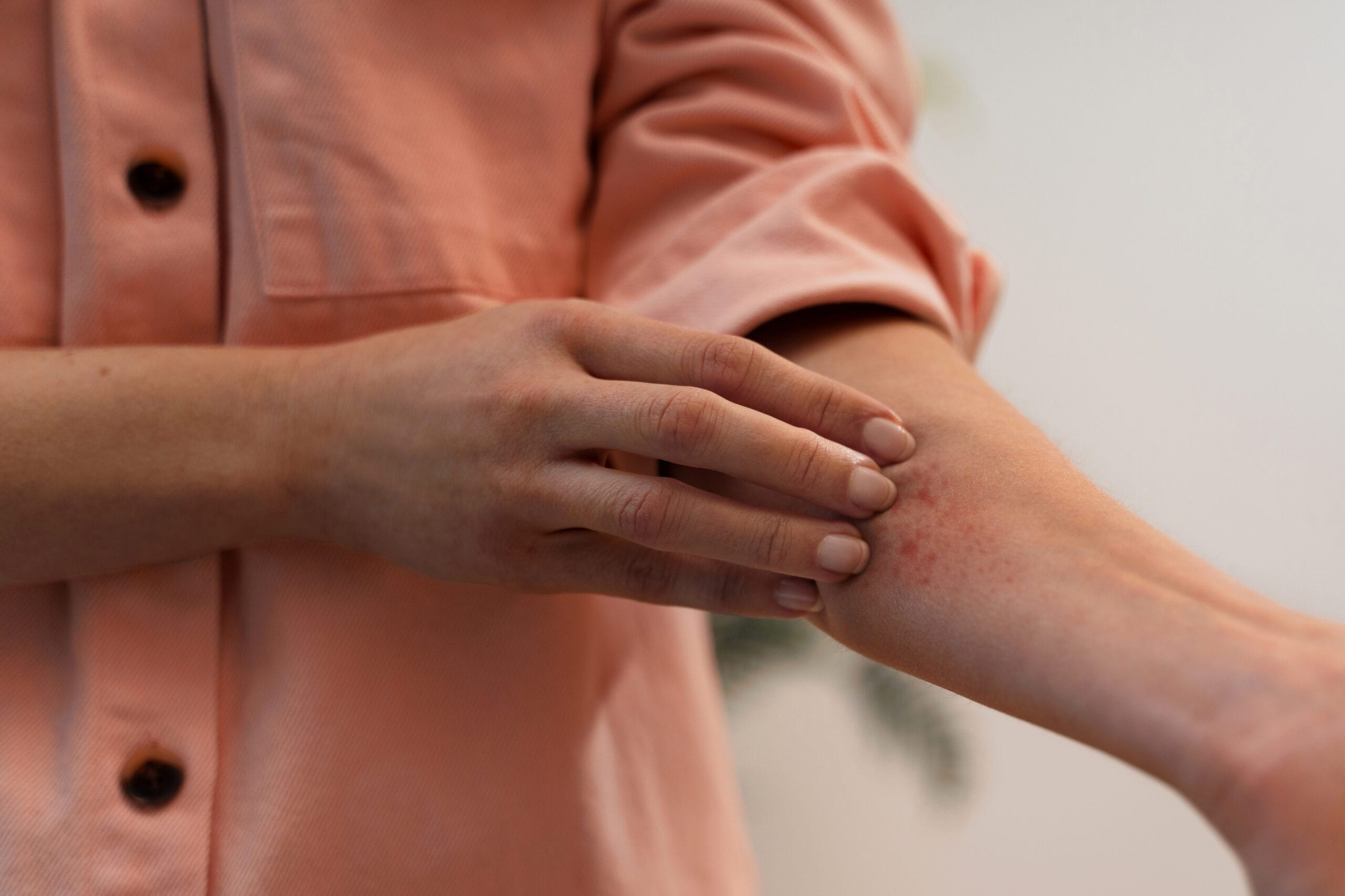

For nearly 2000 years (Celsus and Galenus of Rome), we recognize redness, warmth, swelling, pain, and altered function as immediate cardinal signs of inflammation, the start of an innate immune reaction.
Viewpoint on:
Levine JD, Clark R, Devor M, Helms C, Moskowitz MA, Basbaum AI. Intraneuronal substance P contributes to the severity of experimental arthritis. Science. 1984;226:547-549
Introduction
With the knowledge of Celsus and Galen, in the 19th and early 20th century, several scientists started systematic studies on these cardinal signs of inflammation. In 1876, Salomon Stricker (1834-1898, Vienna) published a paper where he electrically stimulated the dorsal sensory roots of the Nervus ischiadicus. He observed an immediate vasodilation in corresponding areas (1). This was a clear sign of neuronally induced swelling and reddening of skin areas, which was confirmed by William Maddock Bayliss (1860-1924, London). In 1906, Gustav Adolf Spiess (1862-1948, Frankfurt a.M.) (3) reported that inflammation can be blocked when reflexes of sensory nerve fibers were blocked with local anesthetics. He clearly recognized the character of an inflammatory reflex.
In 1910, Alexander Ninian Bruce (1882-1968, Edinburgh) took up the ideas of Spiess and studied the reflex in more detail. He recognized that the reddening reflex was not dependent on the central nervous system but more on local reflexes near the inflammatory area, so-called axon reflexes (4,5). Franz Breslauer (1888-1958, Berlin/New York) saw the clear link between traumatization of a nerve and trophic damage of the tissue because necessary growth signals were missing. The denervated tissue becomes entirely defenseless against daily wear and tear. The inability to dilate blood vessels reactively creates a permanent state of vulnerability (6). In 1936, Thomas Lewis (1881-1945, London) discovered the flare response based on the axon reflex with an involvement of healthy skin and hypersensitivity to pain in a wider area unaffected by the primary stimulus (7). In 1960, Chapman and colleagues found a first polypeptide of the axon reflex called neurokinin that is locally released into blister fluid (8). In the 1960s, Jancso and colleagues extended the concept of neurogenic inflammation with many important studies (9).
With the discovery of substance P in 1931 by Ulf von Euler (10) and the later correct allocation to sensory nerve fibers by Lembeck in 1953 (11) and, particularly, by Otskua and Konishi in 1976 (12), the important role of substance P was found and the platform was prepared to study substance P in the joint.
The authors
Jon Levine (1945- ) received his degrees from the University of Michigan (1966), his PhD from Yale University (1972), and the MD from the University of California in San Francisco (1978). He joined the UCSF faculty in 1987 and became full professor in 1993. His work focused on analgesia and pain as well as arthritis inflammation whereby he looked mainly on the phenomenon of plasma extravasation, a very acute immediate response tightly linked to neurogenic inflammation.
Alan I. Basbaum (1947- ) is a Canadian American medical researcher, and professor and chair of the Department of Anatomy at the University of California in San Francisco. He received his Bachelor of Science at McGill University in Montreal, and his PhD from the University of Pennsylvania, Philadelphia. He holds fellowships with the American Academy of Arts and Sciences and the Royal Society of the United Kingdom, alongside memberships in both the Institute of Medicine and the National Academy of Sciences, the latter of which he was elected to in April 2019. Additionally, he served as the Editor-in-Chief of Pain, the official journal of the International Association for the Study of Pain, from 2003 to 2012.
The results (13)
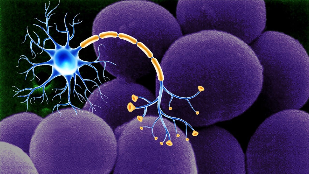
The scientists knew that the important neuropeptide substance P is a neurotransmitter of sensory nerve fibers released from the nerve terminals, and that this neuropeptide is a mediator of neurogenic inflammation. They linked innervation density of substance P – containing nerve fibers with severity of experimental arthritis. They demonstrated that joints with a high density of these nerve fibers (ankle joint in the rat) had more severe arthritis than joints with lower nerve fiber density like the knee joint. Upon injection of substance P into a joint, severity of arthritis was much increased, which linked the neuropeptide to the immediate inflammatory response. At the time of this publication, the exact mechanism by which substance P acts were unknown. Nevertheless, they correctly mentioned “…substance P produces many of the tissue changes of acute inflammation, including vasodilation, increased vascular permeability, pavementing of leukocytes in venules (sticking to the walls), stimulation of phagocytosis by polymorphonuclear leukocytes, and mast cell degranulation.”
Today, we know that substance P has many direct proinflammatory effects like those mentioned above in the context of acute inflammation but also the following ones: stimulation of oxygen radicals from macrophages and neutrophils, cytokine stimulation (IL-1, IL-2, IL-4, IL-6, TNF, IFNg and others), increase of immunoglobulin secretion (IgA, IgG, IgE), stimulation of T and B cell proliferation, support of T helper type 1/2 cells, increase of NFkappaB signaling, and several more. This neuropeptide enhances both arms of the immune system: innate and adaptive.
Neuroimmunomodulation also published papers on substance P and immune function (14-20 and many more).
References
- Stricker S. Untersuchungen über die Gefäßnerven-Wurzeln des Ischiadicus. Sitzungsberichte der Kaiserlichen Akademie der Wissenschaften: Mathematisch-Naturwissenschaftliche Classe 1876;74:173-185
- Bayliss WM. On the origin from the spinal cord of the vaso-dilator fibres of the hind-limb, and on the nature of these fibres. J Physiol. 1901;26:173-209
- Spiess GA. Die Bedeutung der Anästhesie in der Entzüdungstherapie. Münchner Mediz. Wochenschr 1906;53:345-351
- Bruce AN. Über die Beziehung der sensiblen Nervenendigungen zum Entzündungsvorgang. Arch Exp Pathol Pharmakol. 1910;63:424-433
- Bruce AN. Vasodilator axon-reflexes. Q J Exp Physiol. 1913;6:339-354
- Breslauer F. Die Pathogenese des trophischen Gewebeschadens nach der Nervenverletzung. Chir Deut Z. 1919;150:50-81
- Lewis T. Experiments relating to cutaneous hyperalgesia and its spread through somatic nerves. Clin Sci. 1936;2:373-423
- Chapman LF, Ramos A, Goodell H, Wolff HG. Neurokinin – a polypeptide formed during neuronal activity in man. Observations on the axon reflex and antidromic dorsal root stimulation. Trans Am Neurol Assoc. 1960;85:42-45
- Jancso N, Jancso-Gabor A, Szolcsanyi J. Direct evidence for neurogenic inflammation and its prevention by denervation and by pretreatment with capsaicin. Br J Pharmacol Chemother. 1967;31:138-151
- von Euler US, Gaddum JH. An unidentified depressor substance in certain tissue extracts. J Physiol. 1931;72:74-87
- Lembeck F. Central transmission of afferent impulses. III. Incidence and significance of the substance P in the dorsal roots of the spinal cord. Naunyn-Schmiedebergs Archiv fur experimentelle Pathologie und Pharmakologie. 1953;219:197-213
- Otsuka M, Konishi S. Release of substance P-like immunoreactivity from isolated spinal cord of newborn rat. Nature. 1976;264:83-84
- Levine JD, Clark R, Devor M, Helms C, Moskowitz MA, Basbaum AI. Intraneuronal substance P contributes to the severity of experimental arthritis. Science. 1984;226:547-549
- Joachim RA, Sagach V, Quarcoo D, Dinh T, Arck PC, Klapp BF. Upregulation of tumor necrosis factor-alpha by stress and substance p in a murine model of allergic airway inflammation. Neuroimmunomodulation. 2006;13:43-50
- Manske JM, Hanson SE. Substance-P-mediated immunomodulation of tumor growth in a murine model. Neuroimmunomodulation. 2005;12:201-210
- Santoni G, Amantini C, Lucciarini R, Perfumi M, Pompei P, Piccoli M. Neonatal capsaicin treatment affects rat thymocyte proliferation and cell death by modulating substance P and neurokinin-1 receptor expression. Neuroimmunomodulation. 2004;11(3):160-172
- Kang BN, Kim HJ, Jeong KS, Park SJ, Kim SH, Kim SR, Kim TH, Ryu SY. Regulation of leukocyte function-associated antigen 1-mediated adhesion by somatostatin and substance P in mouse spleen cells. Neuroimmunomodulation. 2004;11(2):84-92
- Ruocco HH, Fernandes GA, Namer IJ, Depaulis A, Levy S. Hypothalamic response to experimental allergic encephalomyelitis: role of substance P. Neuroimmunomodulation. 2004;11:28-35
- Santoni G, Amantini C, Lucciarini R, Pompei P, Perfumi M, Nabissi M, Morrone S, Piccoli M. Expression of substance P and its neurokinin-1 receptor on thymocytes: functional relevance in the regulation of thymocyte apoptosis and proliferation. Neuroimmunomodulation. 2002-2003;10:232-246
- Ryu SY, Jeong KS, Yoon WK, Park SJ, Kang BN, Kim SH, Park BK, Cho SW. Somatostatin and substance P induced in vivo by lipopolysaccharide and in peritoneal macrophages stimulated with lipopolysaccharide or interferon-gamma have differential effects on murine cytokine production. Neuroimmunomodulation. 2000;8:25-30
(Featured image declaration: designed by Freepik – Magnific.com)